-
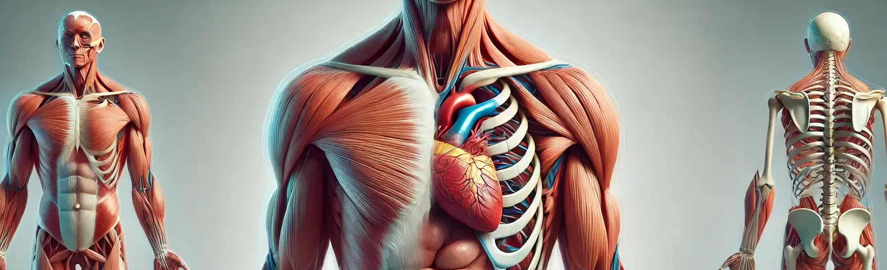
- Muscle tissue is a specialized tissue found in animals which functions by contracting and thereby causing movement.
- This contraction is achieved through the interaction of actin and myosin filaments within muscle cells, known as muscle fibers.
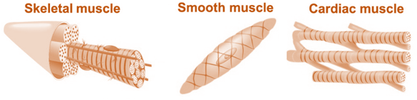
- Muscle tissue is categorized into three types: skeletal muscle, cardiac muscle, and smooth muscle, each with distinct structures and functions.
Types of Muscle Tissue

-
Skeletal Muscle
- Structure: Long, cylindrical, multinucleated fibers with striations from actin and myosin filaments arranged in sarcomeres.
- Location: Attached to bones by tendons.
- Function: Responsible for voluntary movements, posture maintenance, and heat generation.
- Control: Voluntary, via the somatic nervous system.
-
Cardiac Muscle
- Structure: Branched cells (cardiomyocytes) connected by intercalated discs with gap junctions and desmosomes; striated like skeletal muscle.
- Location: Walls of the heart.
- Function: Pumps blood rhythmically and continuously.
- Control: Involuntary, regulated by the autonomic nervous system and intrinsic conduction systems.
-
Smooth Muscle
- Structure: Spindle-shaped cells with a single central nucleus; lacks striations due to less organized actin and myosin.
- Location: Walls of hollow organs (e.g., intestines, blood vessels, bladder, uterus).
- Function: Involuntary movements such as peristalsis, vasoconstriction, and contractions during childbirth.
- Control: Involuntary, via the autonomic nervous system.
Structure of Muscle tissue:
- Skeletal muscle: Multinucleated, long, cylindrical cells with a striated appearance.
- Smooth muscle: Spindle-shaped, uninucleated cells without striations.
- Cardiac muscle: Uninucleated, branched cells with striations and intercalated discs.
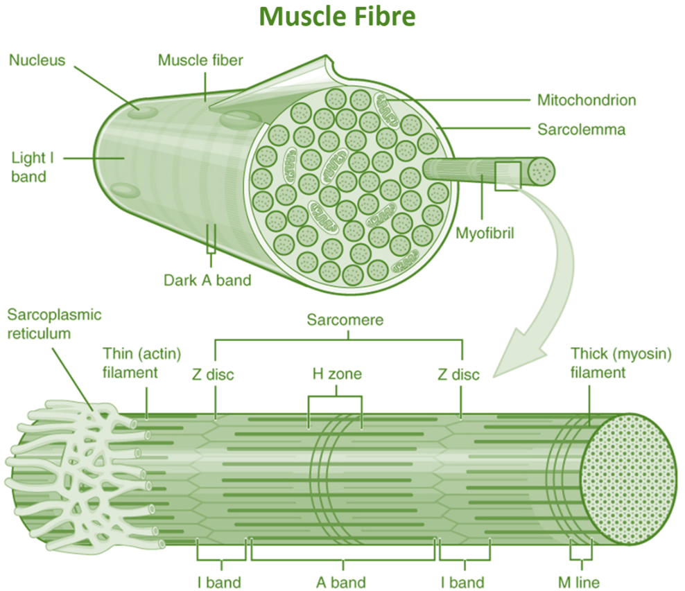
Microscopic Structure

microscopic structure of muscle tissues - Myofibrils:
- Each muscle fiber contains numerous myofibrils, which are the contractile elements made up of repeating units called sarcomeres.
- Sarcomeres:
- The basic functional unit of a myofibril, consisting of actin (thin) and myosin (thick) filaments. The arrangement of these filaments gives skeletal and cardiac muscle their striated appearance.
- Sarcoplasmic Reticulum:
- A specialized form of the endoplasmic reticulum that stores and releases calcium ions, which are essential for muscle contraction.
- Mitochondria:
- Muscle cells contain numerous mitochondria to meet the high energy demands required for contraction.
Location:
- Skeletal muscle: Attached to bones by tendons.
- Smooth muscle: Walls of internal organs like the stomach, intestines, and blood vessels.
- Cardiac muscle:
Functions:
- Movement: Muscle contractions enable body movement, facial expressions, and posture.
- Regulation: Smooth muscle contractions regulate blood flow and control the passage of food through the digestive system.
Pumping: Cardiac muscle contractions pump blood throughout the body.
Thank you for reading from Firsthope's notes, don't forget to check YouTube videos!