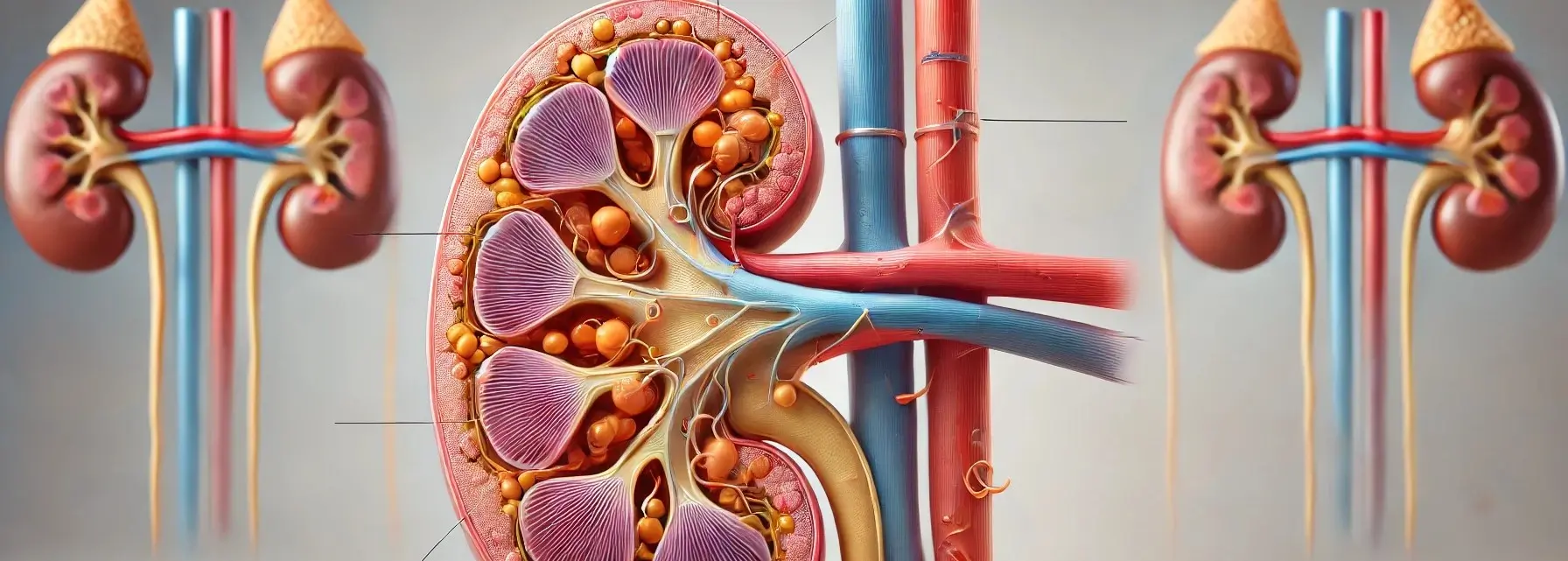
- Nephron anatomy are the microscopic structural and functional units of the kidney, crucial for the process of filtering blood, removing waste, and regulating fluid and electrolyte balance.
- Each kidney contains about 1 million nephrons.
- A nephron is composed of two main parts: the renal corpuscle and the renal tubule.

Demo Ad
This is a sample ad placement!
This is a sample ad placement!
Renal Corpuscle
- The renal corpuscle is the initial filtering component of the nephron and is located in the renal cortex.
- It consists of two main structures:
-
Glomerulus:
- A cluster of capillaries where blood filtration begins, separating water and small solutes from larger molecules.
-
Bowman’s Capsule:
- Encases the glomerulus, with an inner layer (podocytes) and an outer layer, collecting the filtrate to pass it to the renal tubule.
-
Renal Tubule
- The renal tubule is a long, coiled tube that converts the filtrate from the Bowman’s capsule into urine by reabsorption and secretion.
- It is divided into several segments:
-
Proximal Convoluted Tubule (PCT):
- Reabsorbs water, sodium, glucose, and nutrients from the filtrate.
-
Loop of Henle:
- Extends into the medulla, with descending and ascending limbs to concentrate urine by regulating water and salt levels.
-
Distal Convoluted Tubule (DCT):
- Further adjusts ion balance and pH, playing a key role in regulating potassium, sodium, and calcium.
-
Collecting Duct:
- Receives processed filtrate from multiple DCTs, fine-tuning water reabsorption and determining urine’s final concentration and volume.
-
Demo Ad
This is a sample ad placement!
This is a sample ad placement!
Thank you for reading from Firsthope's notes, don't forget to check YouTube videos!