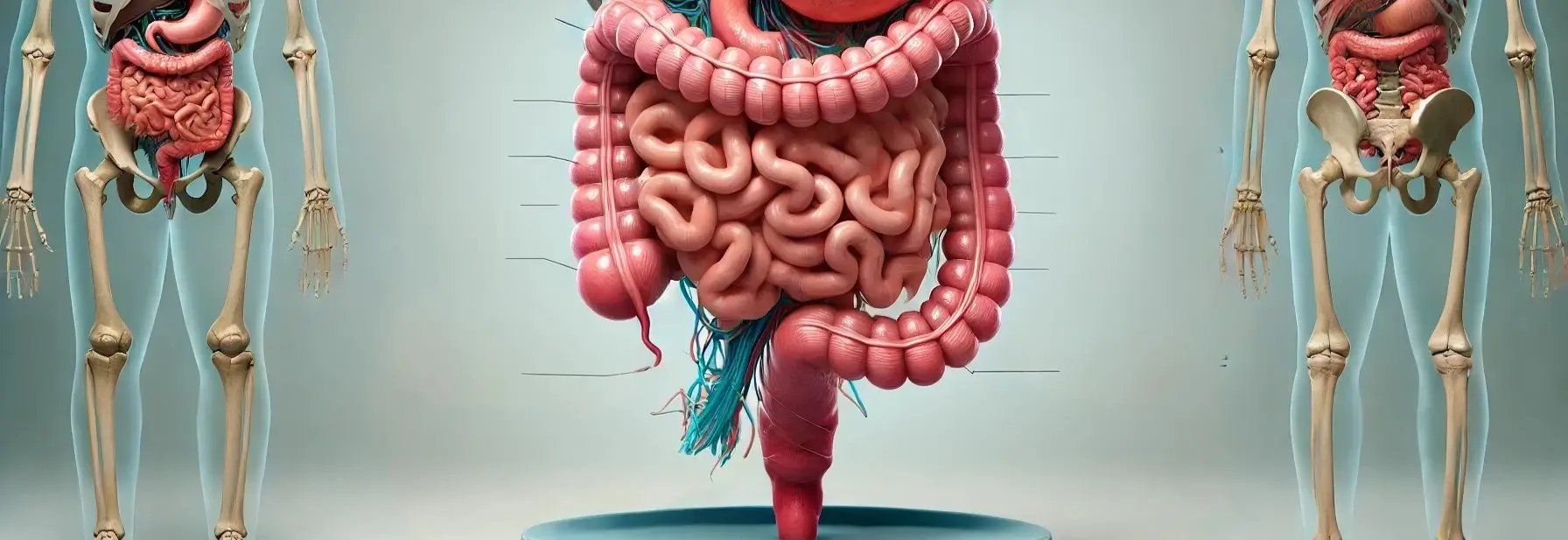
- The Anatomy of GI Tract, also known as the digestive tract, is a continuous tube that runs from the mouth to the anus.
- It is responsible for breaking down food, absorbing nutrients, and eliminating waste.
Here’s a brief overview of the four primary layers of the gastrointestinal tract (GIT) The innermost layer responsible for secretion, absorption, and protection. from the innermost to the outermost layer:
Advertisements
1. Mucosa:
- It has three sub-layers: epithelium, lamina propria, and muscularis mucosae.
Advertisements
2. Submucosa:
- A layer of connective tissue that contains blood vessels, lymphatic vessels, nerves, and glands.
3. Muscularis externa:
- A layer of smooth muscle that propels food through the Anatomy of GI Tract.
- It has an inner circular layer, an outer longitudinal layer, and an additional oblique layer in the stomach.
Advertisements
4. Serosa/Adventitia:
- The outermost layer, composed of connective tissue and mesothelium (serosa) or connective tissue (adventitia) that anchors the GIT to surrounding structures.

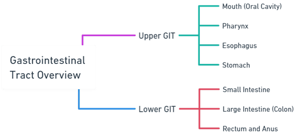
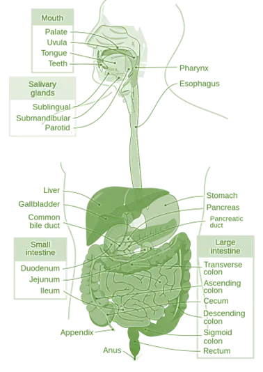
Upper GI Tract
1. Mouth (Oral Cavity):
- Begins the digestive process.
- Structures include the lips, cheeks, palate, tongue, teeth, and salivary glands.
- Salivary glands (parotid, submandibular, and sublingual) produce saliva that moistens food and begins the digestion of carbohydrates with the enzyme amylase.
2. Pharynx:
- A muscular tube that connects the mouth to the esophagus.
- Plays a role in swallowing, directing food from the mouth to the esophagus, and preventing food from entering the trachea (windpipe) through the epiglottis.
Advertisements
3. Esophagus:
- A muscular tube about 10 inches long, running from the pharynx to the stomach.
- Transports food to the stomach via peristaltic movements.
- The lower esophageal sphincter (LES) controls the passage of food into the stomach and prevents the backflow of gastric contents.
4. Stomach:
- A J-shaped muscular organ that stores, mixes, and digests food.
- Divided into four regions: cardia, fundus, body (corpus), and pylorus.
- The stomach lining contains gastric glands that secrete gastric juice, including hydrochloric acid and digestive enzymes like pepsin.
Lower GI Tract
Small Intestine:
- Approximately 20 feet long, divided into three sections: duodenum, jejunum, and ileum.
- The duodenum receives chyme from the stomach, along with bile from the liver and gallbladder, and pancreatic juices from the pancreas, which aid in digestion.
- The jejunum and ileum are mainly involved in the absorption of nutrients and water.
Advertisements
Large Intestine (Colon):
- About 5 feet long, divided into the cecum, ascending, transverse, descending, and sigmoid colon, and the rectum.
- Absorbs remaining water and electrolytes from indigestible food matter and forms solid waste (feces).
- The colon houses a large number of bacteria that play a role in the fermentation of undigested food, vitamin production, and protection against harmful bacteria.
Rectum and Anus:
- The rectum stores feces until they are expelled from the body.
- The anus is the final part of the GI tract, consisting of internal and external sphincters that control the expulsion of feces.
Accessory Digestive Organs
- In addition to the primary GI tract, several accessory organs contribute to the digestive process:
Advertisements
Salivary Glands:
- As mentioned, these include the parotid, submandibular, and sublingual glands, which secrete saliva to initiate the digestion of carbohydrates.
Liver:
- The body’s largest gland, playing a crucial role in metabolism, detoxification, and the production of bile, which helps in the digestion and absorption of fats.
Gallbladder:
- Stores and concentrates bile from the liver, releasing it into the duodenum to aid in digestion.
Advertisements
Pancreas:
- Produces pancreatic juice containing digestive enzymes and bicarbonate ions, which neutralize the acidity of chyme entering the duodenum from the stomach.