- The eye is a complex sensory organ responsible for detecting light and converting it into electrical signals that the brain can interpret as visual information.
- The human eye has several components that work together to facilitate the process of vision.
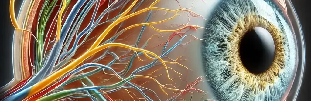
Structure of the Eye
The human eye is a complex organ responsible for vision, made up of several parts: 
- Cornea: The transparent outermost layer that refracts light entering the eye.
- Sclera: The white part of the eye that provides structural support and protection.
- Iris: The colored part of the eye that controls the size of the pupil, regulating the amount of light that enters.
- Pupil: The black circular opening in the center of the iris that allows light to pass through.
- Lens: A transparent, flexible structure that focuses light onto the retina.
- Retina: The innermost layer that contains photoreceptor cells (rods and cones) responsible for detecting light.
- Rods: Photoreceptors that are sensitive to low light levels, crucial for night vision.
- Cones: Photoreceptors responsible for color vision and sharpness of vision in well-lit conditions.
- Optic Nerve: Transmits visual information from the retina to the brain.
- Choroid: The vascular layer of the eye that supplies blood to the retina.
- Macula: The central part of the retina responsible for detailed central vision.
- Fovea: A small depression in the macula that contains a high concentration of cones and is the area of sharpest vision.
- Aqueous Humor: A clear fluid that fills the space between the cornea and the lens, providing nutrients and maintaining intraocular pressure.
- Vitreous Humor: A gel-like substance that fills the space between the lens and the retina, maintaining the shape of the eye
Mechanism of Vision

Advertisements
- Light Entry: Light enters the eye through the cornea, which bends (refracts) the light.
- Pupil Regulation: The iris adjusts the size of the pupil to control how much light enters the eye.
- Focusing: The lens, with the help of ciliary muscles, adjusts its shape (accommodation) to focus light on the retina.
- Image Formation: Light is focused onto the retina, where photoreceptor cells (rods and cones) detect the light and convert it into electrical signals.
- Signal Transmission: These electrical signals are sent to the brain via the optic nerve.
- Image Processing: The brain processes these signals, interpreting them into the images we see.
Accommodation:
- Accommodation refers to the eye’s ability to change the shape of the lens to focus on objects at varying distances.
- When focusing on distant objects, the ciliary muscles relax, and the lens becomes flatter.
- When focusing on close objects, the ciliary muscles contract, making the lens thicker.
- This process allows the eye to maintain a clear image (focus) on the retina regardless of whether the object is near or far.
Diseases of the Eye:
- Cataracts: Clouding of the lens that leads to blurred vision. It often occurs with aging.
- Glaucoma: A condition where increased intraocular pressure damages the optic nerve, leading to vision loss.
- Macular Degeneration: A disease that affects the macula, leading to loss of central vision, common in older adults.
- Myopia (Nearsightedness): A condition where distant objects appear blurry because the light focuses in front of the retina.
- Hyperopia (Farsightedness): A condition where near objects appear blurry because light focuses behind the retina.
- Astigmatism: Irregular curvature of the cornea or lens causing distorted or blurred vision.
- Conjunctivitis (Pink Eye): Inflammation of the conjunctiva, the membrane covering the white of the eye, often caused by infection or allergy.
- Retinal Detachment: Separation of the retina from the underlying tissue, which can lead to blindness if not treated promptly.
Functions:
- Vision: The primary function of the eye is to detect light and convert it into neural signals that are processed by the brain to create images.
- Color Detection: The cones in the retina allow for the perception of different colors.
- Light Intensity Regulation: The iris regulates the size of the pupil to control the amount of light entering the eye, preventing damage to the retina.
- Focus Adjustment (Accommodation): The lens adjusts to focus light precisely on the retina, allowing for clear vision of objects at various distances.
- Depth Perception: The eye contributes to spatial awareness and depth perception, especially when working together with the other eye (binocular vision).
Click Here to Watch the Best Pharma Videos
Advertisements