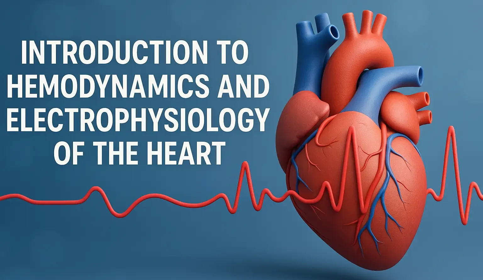
- Introduction to Hemodynamics and Electrophysiology of the Heart: Covers blood flow and cardiac electrical activity.
- Introduction to Hemodynamics and Electrophysiology of the Heart: Key to understanding cardiac output and rhythm regulation.
Hemodynamics
Definition
- Hemodynamics refers to the dynamics of blood flow, including how the heart pumps blood, how blood pressure is generated and regulated, and how blood circulates through the vasculature.
Advertisements
Key Terms
- Cardiac Output (CO) = Heart Rate (HR) × Stroke Volume (SV)
- Stroke Volume (SV): The amount of blood ejected from the ventricle with each heartbeat.
- Preload: The degree of ventricular filling (end-diastolic volume) that stretches the cardiac muscle fibers before contraction.
- Afterload: The resistance the left ventricle must overcome to pump blood into the systemic circulation.
- Contractility: The intrinsic ability of cardiac muscle fibers to contract, independent of preload and afterload.
- Ejection Fraction (EF): The fraction of the end-diastolic volume ejected during systole (SV ÷ EDV).
Regulation of Blood Pressure
- Baroreceptor Reflex: Stretch receptors in the carotid sinus and aortic arch sense changes in blood pressure and modulate sympathetic/parasympathetic outflow.
- Renin-Angiotensin-Aldosterone System (RAAS): Renin (kidneys) → Angiotensin I → Angiotensin II (via ACE in lungs) → Aldosterone (adrenal cortex) → Na+^++/H2_22O retention → ↑ Blood volume and pressure.
- Autonomic Nervous System: Sympathetic stimulation increases heart rate, contractility, and vasoconstriction; parasympathetic stimulation (vagus nerve) decreases heart rate.
Advertisements
Electrophysiology of the Heart
Cardiac Conduction System
- Sinoatrial (SA) Node: The primary pacemaker (60-100 bpm).
- Atrioventricular (AV) Node: Delays conduction from the atria to the ventricles.
- Bundle of His → Purkinje Fibers: Rapid conduction to the ventricles.
Action Potential Phases (Ventricular Myocytes)
- Phase 0 (Depolarization): Rapid influx of Na+^++ → Sharp upstroke.
- Phase 1 (Initial Repolarization): Inactivation of Na+^++ channels; K+^++ efflux begins.
- Phase 2 (Plateau): Balance between Ca2+^{2+}2+ influx (through L-type Ca2+^{2+}2+ channels) and K+^++ efflux.
- Phase 3 (Repolarization): Increased K+^++ efflux; Ca2+^{2+}2+ channels inactivate.
- Phase 4 (Resting Membrane Potential): High K+^++ permeability through K+^++ channels, restoring the resting potential.
Advertisements
Nodal Cells (SA/AV)
- Spontaneous Depolarization (Pacemaker Potential): Slower rise, mainly involving Ca2+^{2+}2+ and reduced K+^++ permeability.
- Autonomic Influences:
- Sympathetic: Increases the slope of pacemaker potential → Increased heart rate.
- Parasympathetic: Decreases slope → Reduced heart rate.

