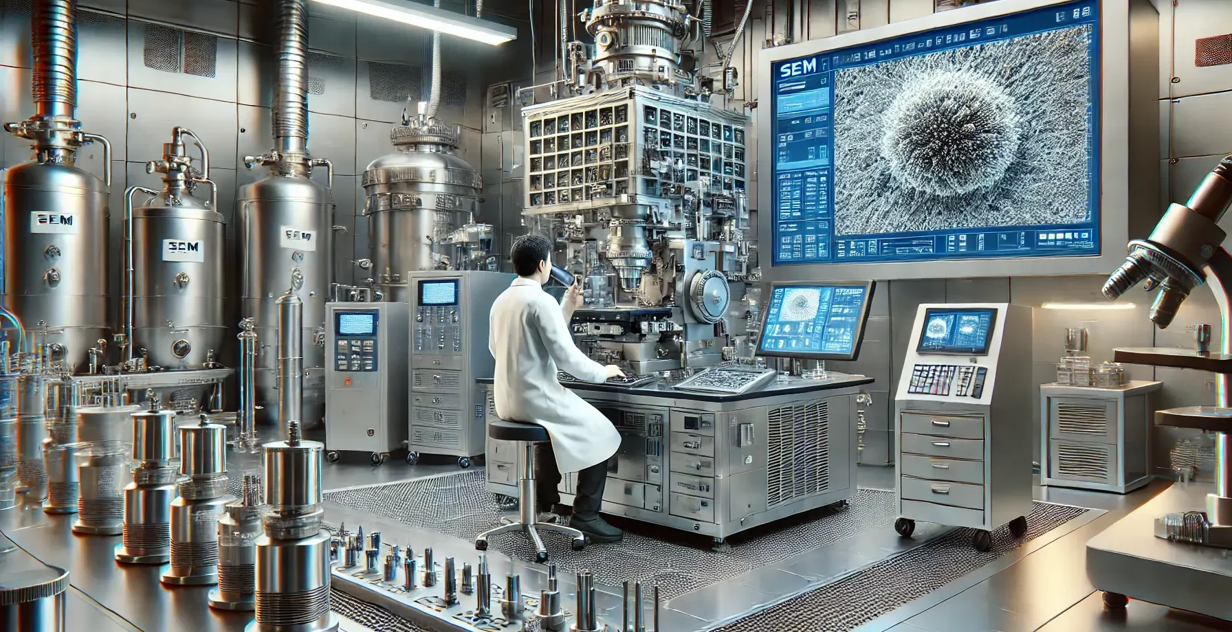
Principle of Scanning Electron Microscopy (SEM)
- SEM scans a focused electron beam across the specimen’s surface. Secondary electrons emitted from the specimen surface are collected to form an image.
- SEM provides high-resolution, three-dimensional images that reveal the specimen’s surface topology.
Procedure for Scanning Electron Microscopy (SEM)
-
Specimen Preparation:
- Fixation: The specimen is fixed to preserve its structure, similar to TEM.
- Dehydration: Critical point drying is used to prevent the collapse of structures caused by surface tension.
- Mounting: The specimen is attached to an aluminum stub using conductive adhesives.
- Coating: A thin layer of conductive metal (e.g., gold or platinum) is sputter-coated on the specimen to prevent charging and improve signal quality.
-
Operation of the SEM:
- Vacuum System: The specimen chamber is evacuated to reduce electron scattering.
- Electron Source: An electron beam is generated using a thermionic or field emission gun.
- Beam Focusing: Electromagnetic lenses are used to focus the electron beam into a fine spot.
- Scanning: The electron beam is raster-scanned over the specimen’s surface.
-
Detection and Imaging:
- Secondary Electron Detector: Secondary electrons emitted from the specimen are collected.
- Image Formation: The intensity of these electrons is used to form a grayscale image displayed on a monitor.
-
Image Processing:
- Adjustments: Brightness, contrast, and focus are modified for optimal image quality.
- Data Capture: Images are saved digitally for further analysis.
Advertisements
Applications
- Surface Morphology: Studying the texture and topography of materials.
- Biology: Examining surface structures of cells and tissues.
- Nanotechnology: Visualizing nanoparticles and nanostructures.
Advantages of Scanning Electron Microscopy:
- High-resolution imaging of surfaces.
- Depth of field allows for 3D-like visualization.
- No need for ultra-thin specimens.
Advertisements
Limitations of Scanning Electron Microscopy:
- Internal structures cannot be viewed.
- Non-conductive specimens require a conductive coating.
- Specimens must withstand vacuum conditions.
Click Here to Watch the Best Pharma Videos
Advertisements